Nikon’s Small World Competition has yet again revealed the concealed marvels of our world via the microscope’s lens. In 2023, this esteemed contest carried on its legacy of honoring the artistic and scientific value of microscopy.
This year’s victorious captures are a tribute to the astonishing allure and intricacy that dwell beneath the surface. Ranging from lively biological specimens to enthralling chemical formations, these images exhibit the varied and hypnotizing realm of microphotography.
Hassanain Qambari, 1st place
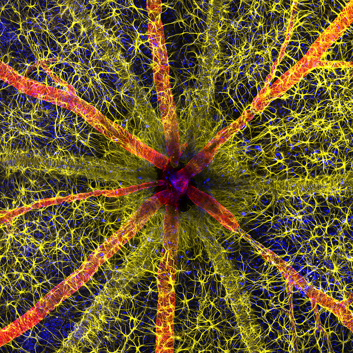
“Rodent optic nerve head showing astrocytes, contractile proteins and retinal vasculature”
Ole Bielfeldt, 2nd place
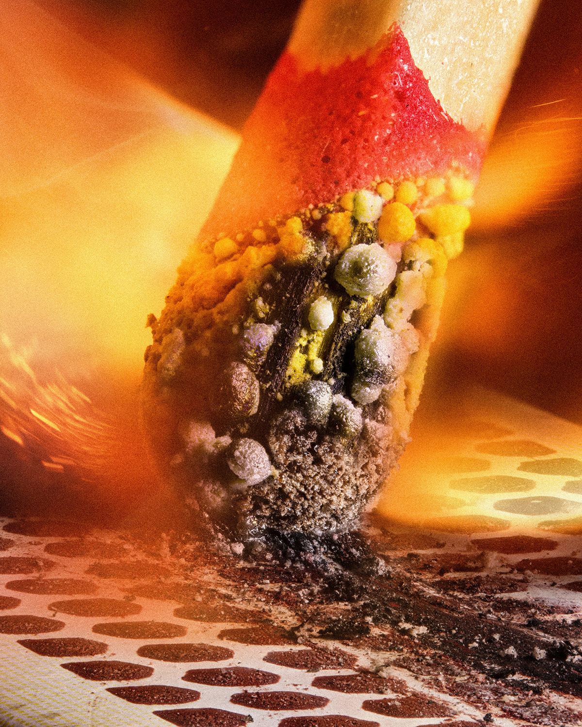
Match igniting on the friction surface of the box”
Malgorzata Lisowska, 3rd place
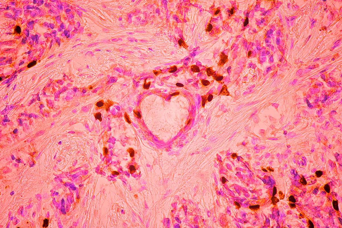
“Breast cancer cells”
John-Oliver Dum, 4th place
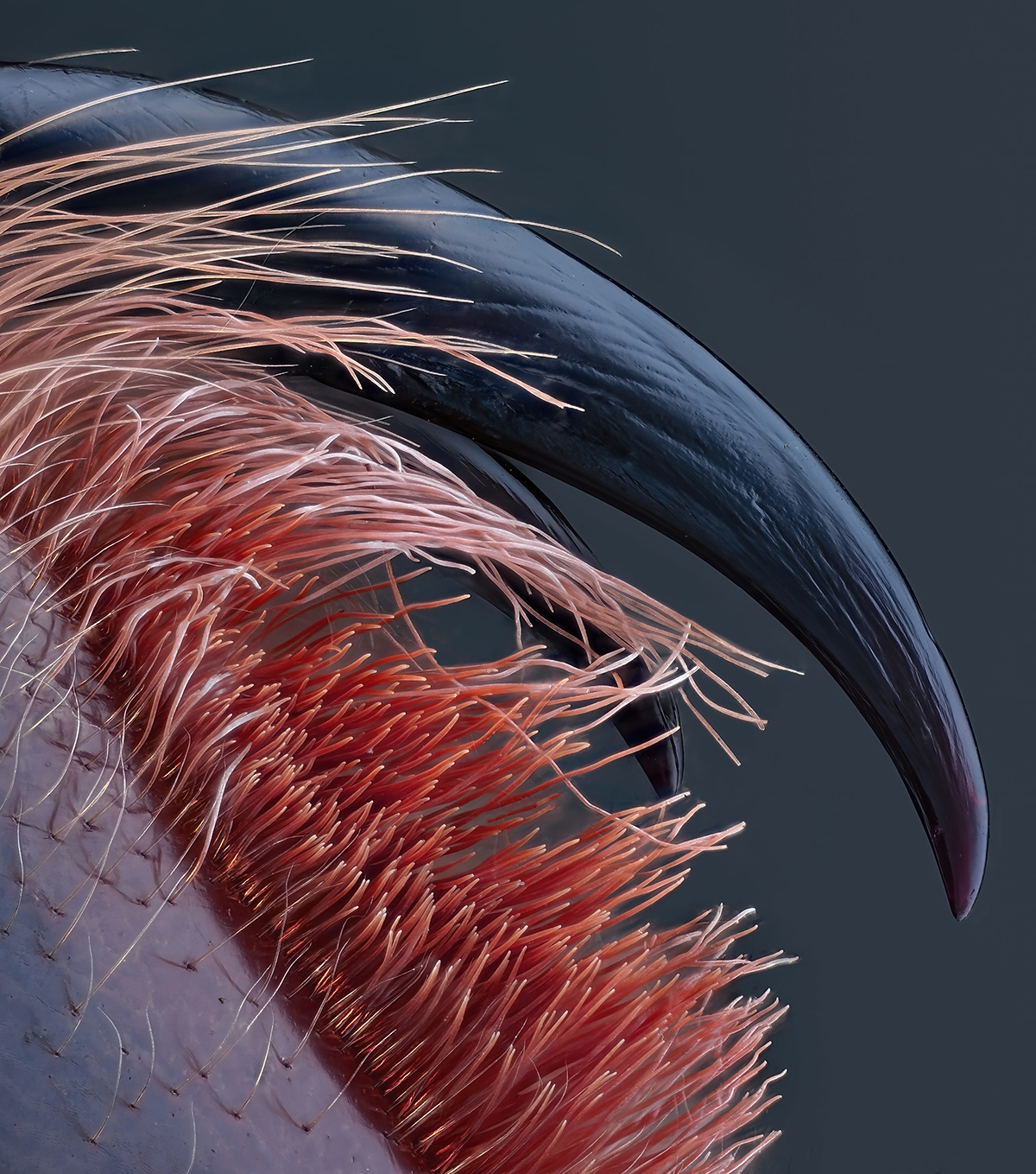
“Poisonous fangs of a small tarantula”
Dr. David Maitland, 5th place
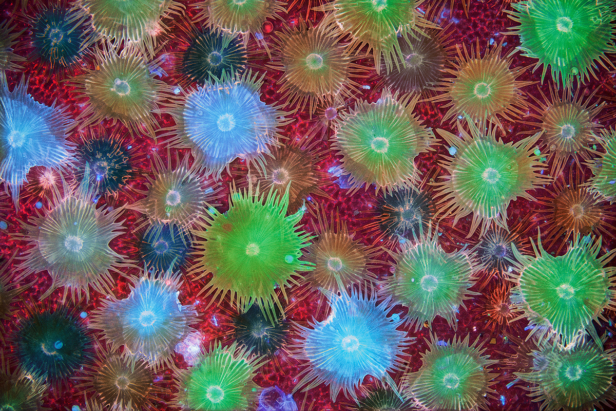
“Auto-fluorescent defensive hairs covering the surface of Eleagnus angustifolia leaves exposed to UV rays”
Timothy Boomer, 6th place
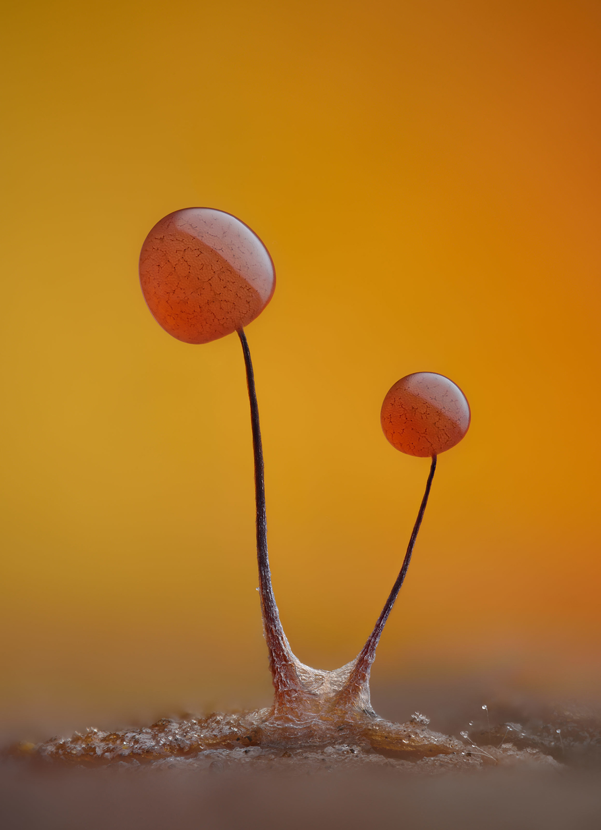
“Slime mold showing hair fibers through its translucent peridium”
Dr. Grigorii Timin, 7th place

“Mouse embryo”
Stefan Eberhard, 8th place
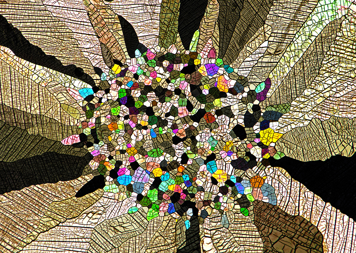
“Caffeine crystals”
Vaibhav Deshmukh, 9th place
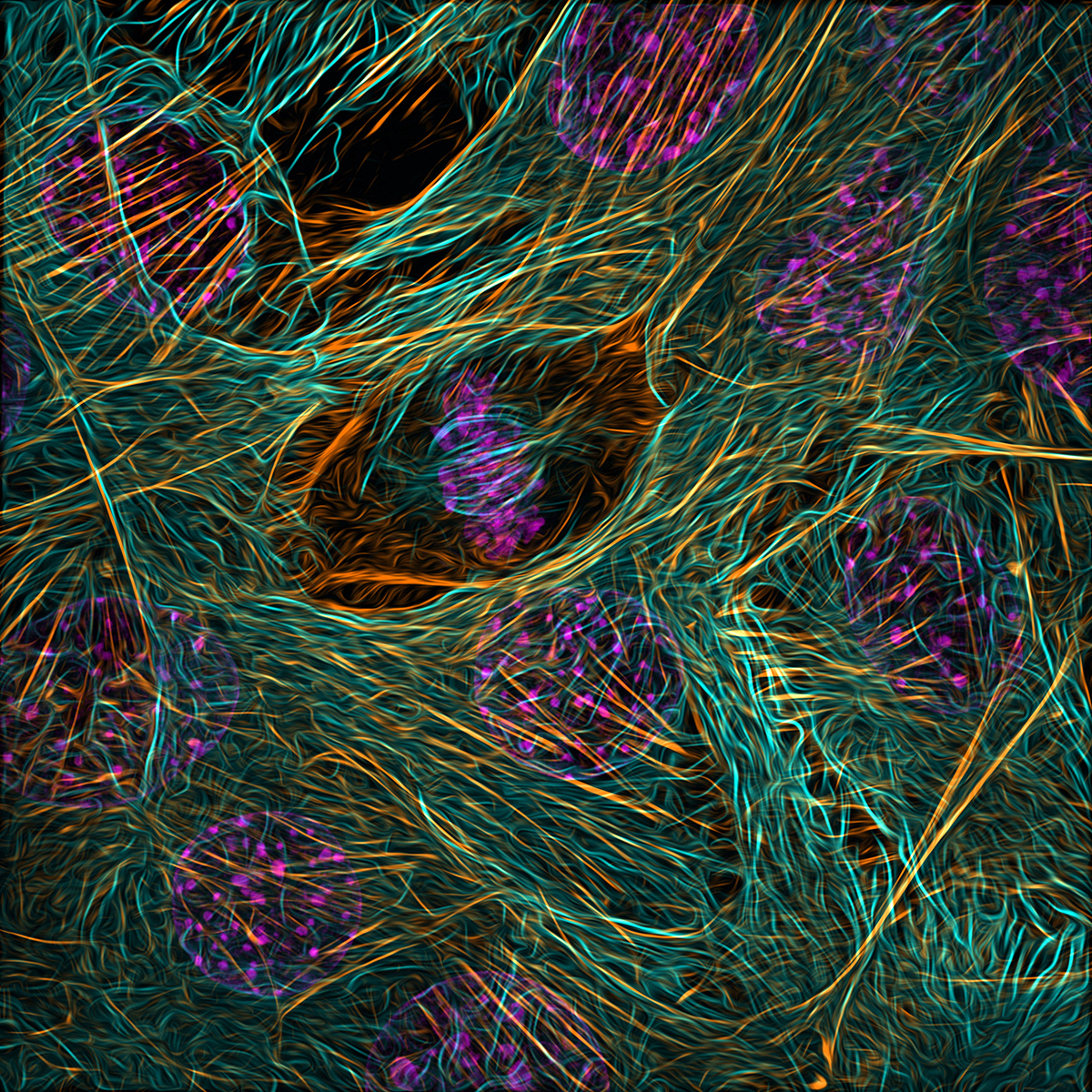
“ Cytoskeleton of a dividing myoblast; tubulin, F-actin and nucleus “
Melinda Beccari, 10th place

“Motor neurons cultured in a microfluidic device for separation of cell bodies and axons”
Dr. Diego García, 11th place

“Crystallized sugar syrup”
Sherif Abdallah Ahmed, 12th place
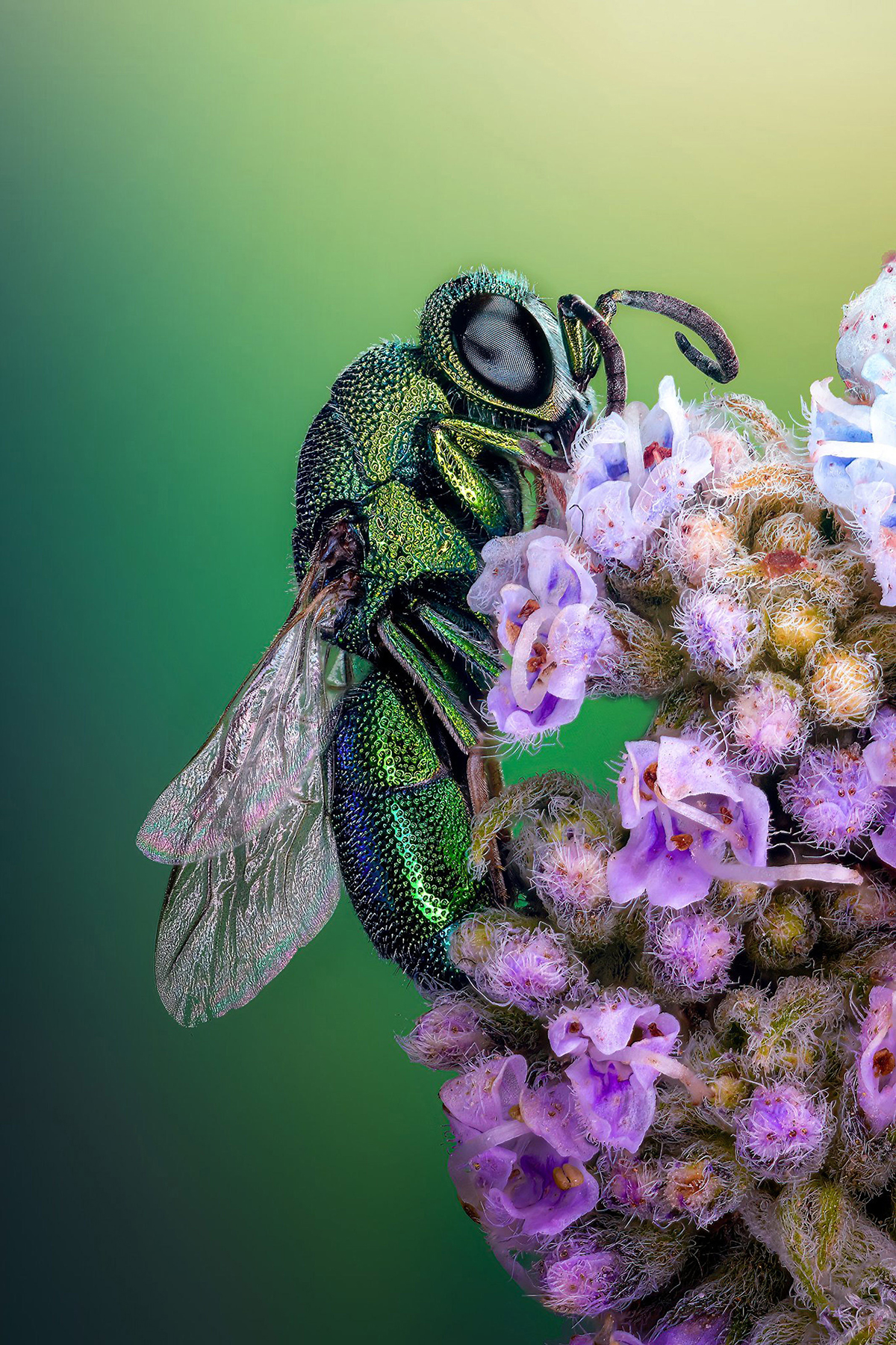
“Cuckoo wasp standing on a flower”
Satu Pavonalo, 13th place

“Blood and lymphatic vascular systems in the ear skin of an adult mouse”
John-Oliver Dum, 14th place

“Sunflower pollen on an acupuncture needle”
Dr. Pichaya Lertvilai, 15th place
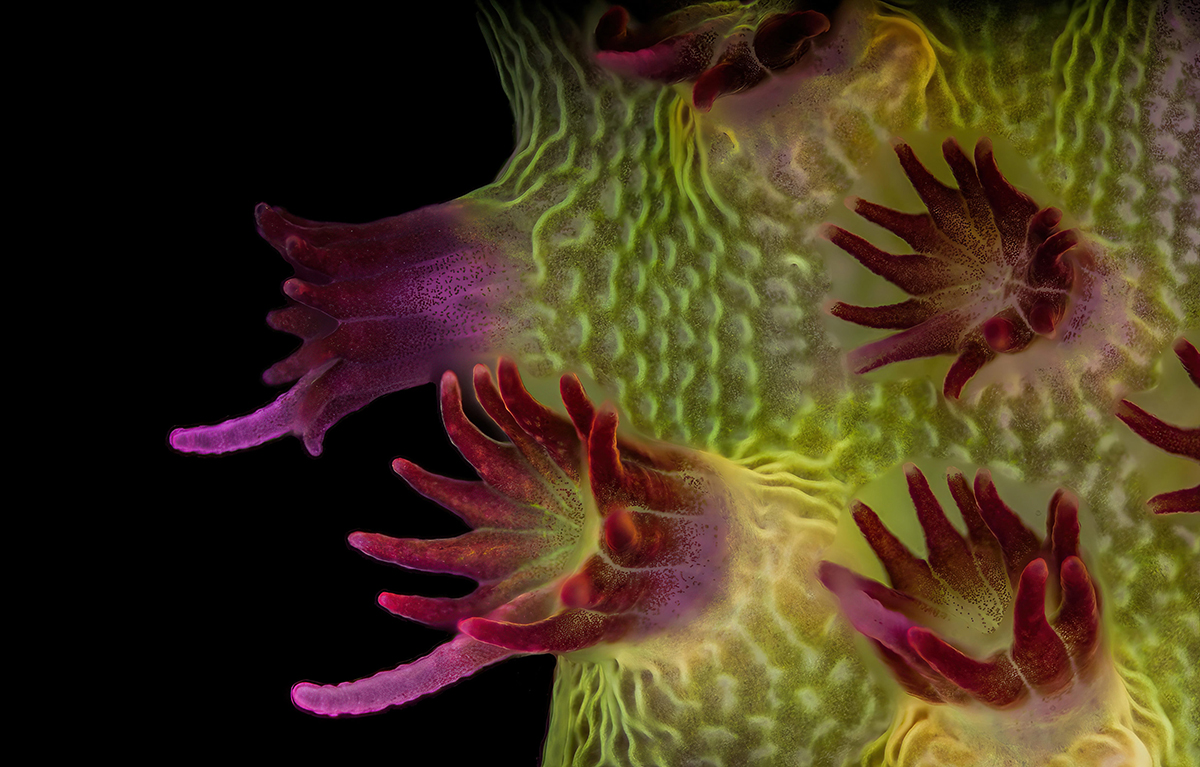
“Fluorescent image of an Acropora sp. showing individual polyps with symbiotic zooxanthellae”
Dr. Diego García, 16th place
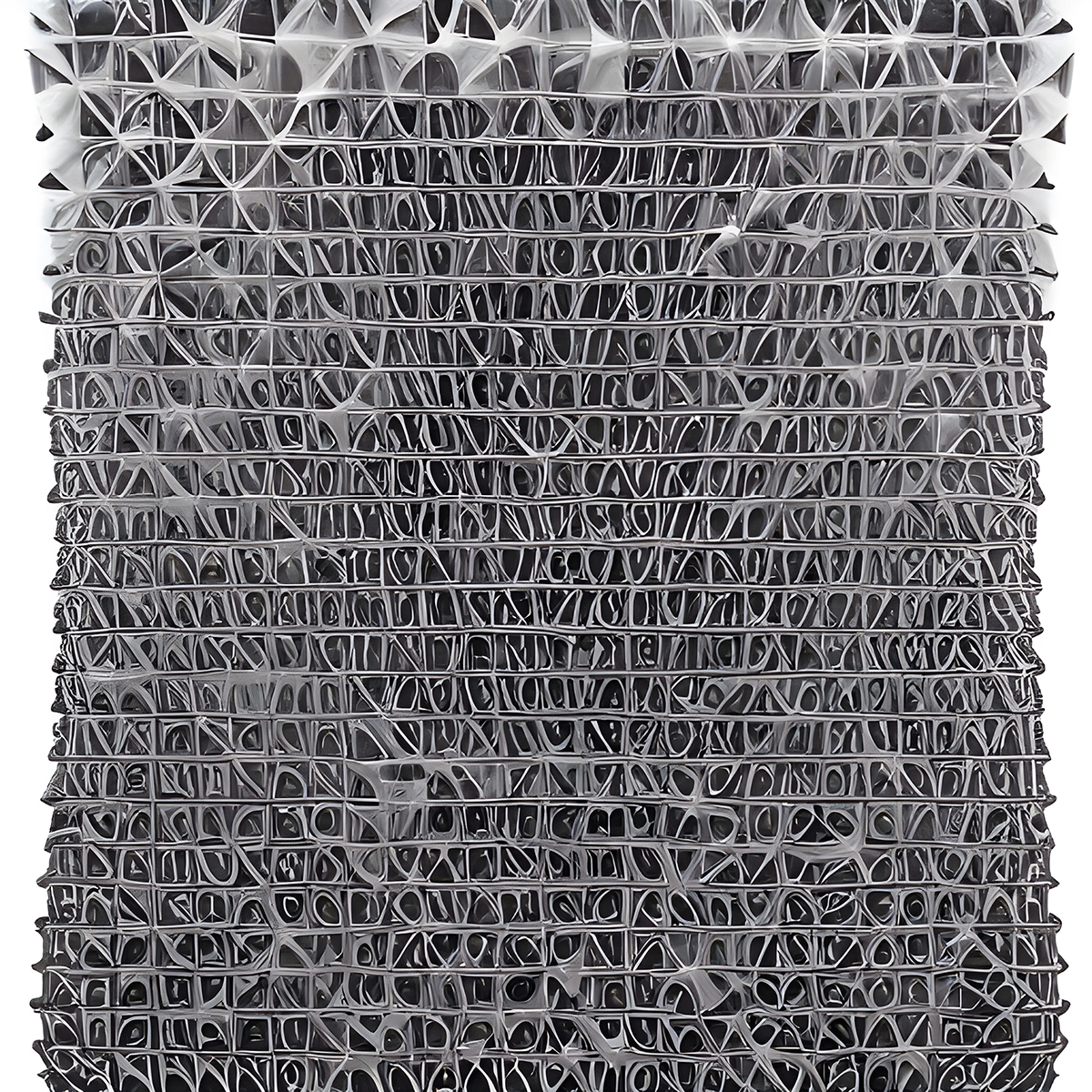
“Carbon nanotubes”
Yuan Ji, 17th place
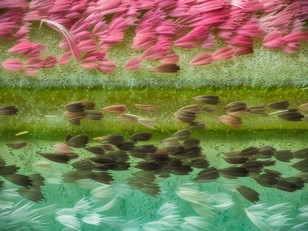
“Chinese Moon Moth”
Scott Peterson, 18th place
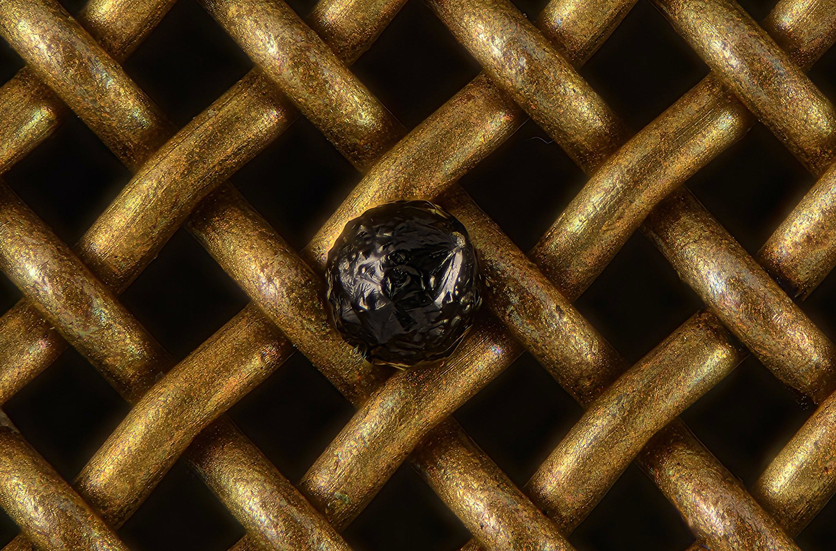
“ A cryptocrystalline micrometeorite resting on a No. 80 test sieve ”
Marek Miś, 19th place
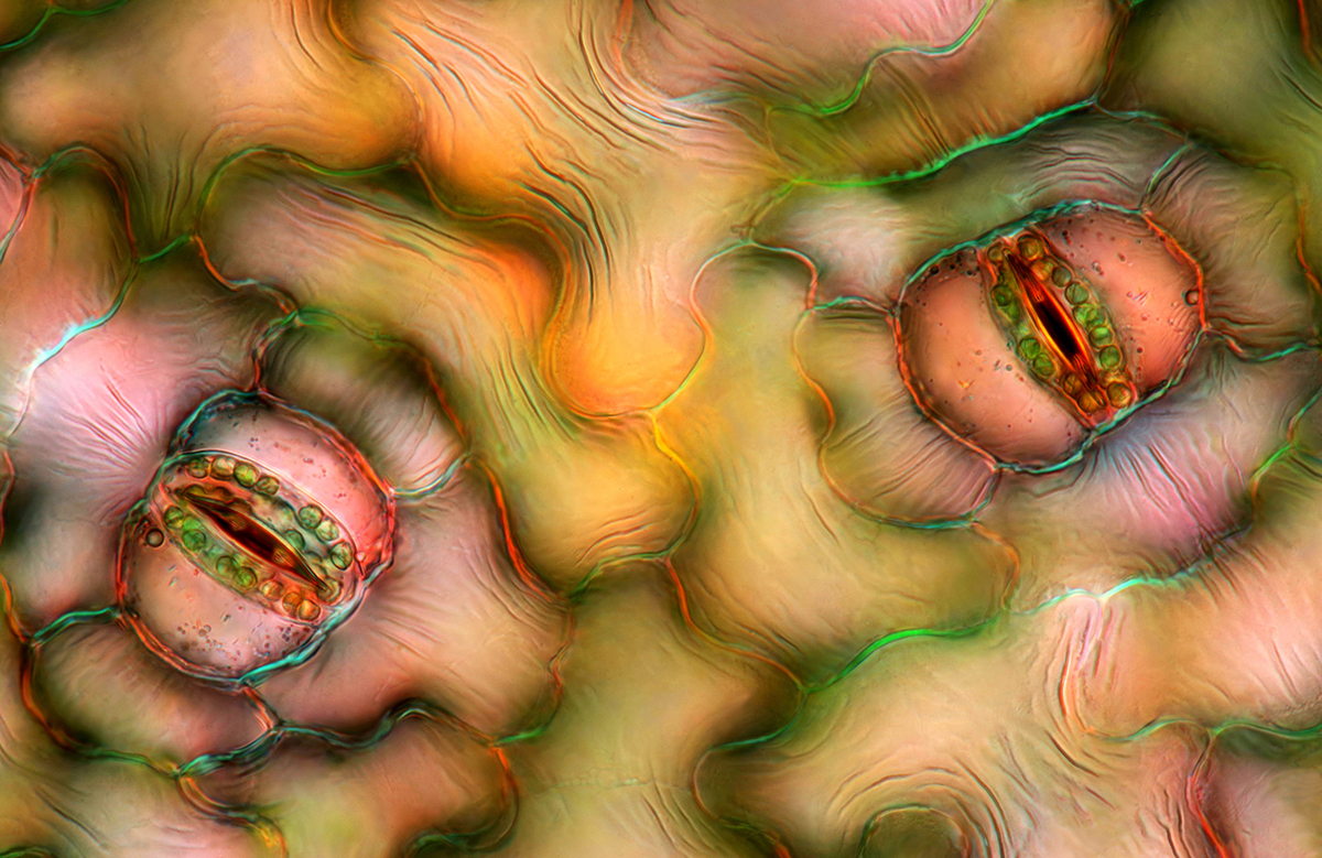
“Peace Lily Stomata”
Daniel Castranova, 20th place
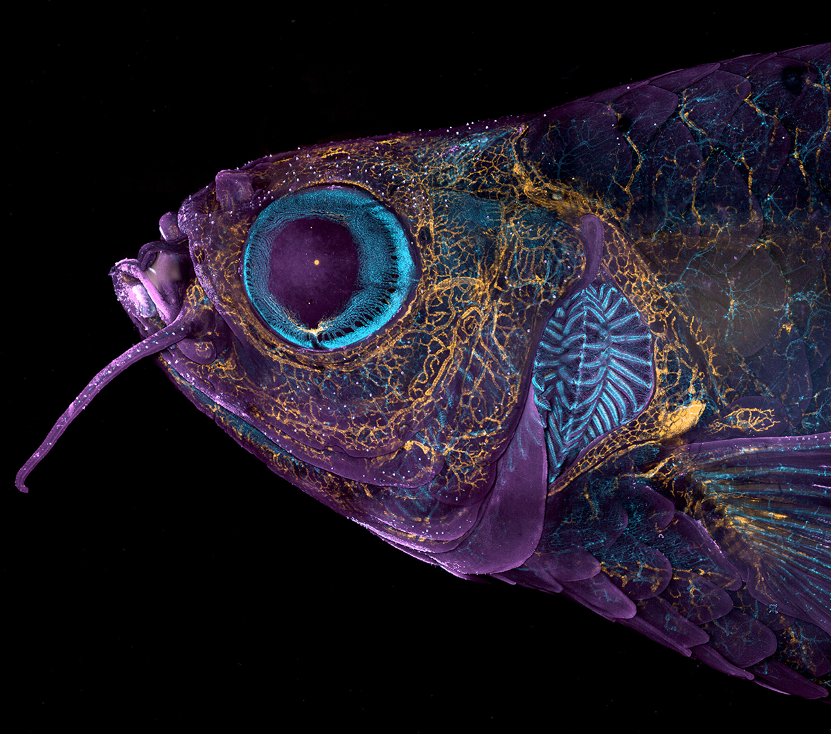
“ Adult transgenic zebrafish head showing blood vessels (blue), lymphatic vessels (yellow), and skin and scales (magenta) ”
 Barnorama All Fun In The Barn